Imagine a medical landscape where cellular decay isn’t permanent and placing human organs in a state of suspended animation is an everyday reality. For decades, the formation of destructive ice crystals made freezing delicate biological tissues impossible. However, specialists have recently demonstrated that neural tissue can completely regain its functional capabilities even after plunging to -196°C. This remarkable breakthrough fundamentally shifts our understanding of cognitive preservation and transplant medicine.
A dedicated research team at the Friedrich-Alexander University Erlangen-Nuremberg has accomplished what many deemed pure science fiction. In an unprecedented milestone, adult mouse brain tissue successfully resumed its complex activity following a specialized vitrification process. The findings confirm that the hippocampus—the brain’s primary engine for memory and learning—can survive extreme freezing for several days. Once warmed, this critical region seamlessly went back to transmitting electrical impulses across its neural networks.
The Science Behind Vitrification
To truly appreciate this medical milestone, we have to look at the primary obstacle: freezing water. When biological material is preserved using conventional cooling methods, internal moisture transforms into jagged ice crystals that shred cell walls and sever delicate neural connections. It is essentially like placing a running computer in a deep freeze until expanding ice snaps every circuit on the motherboard.
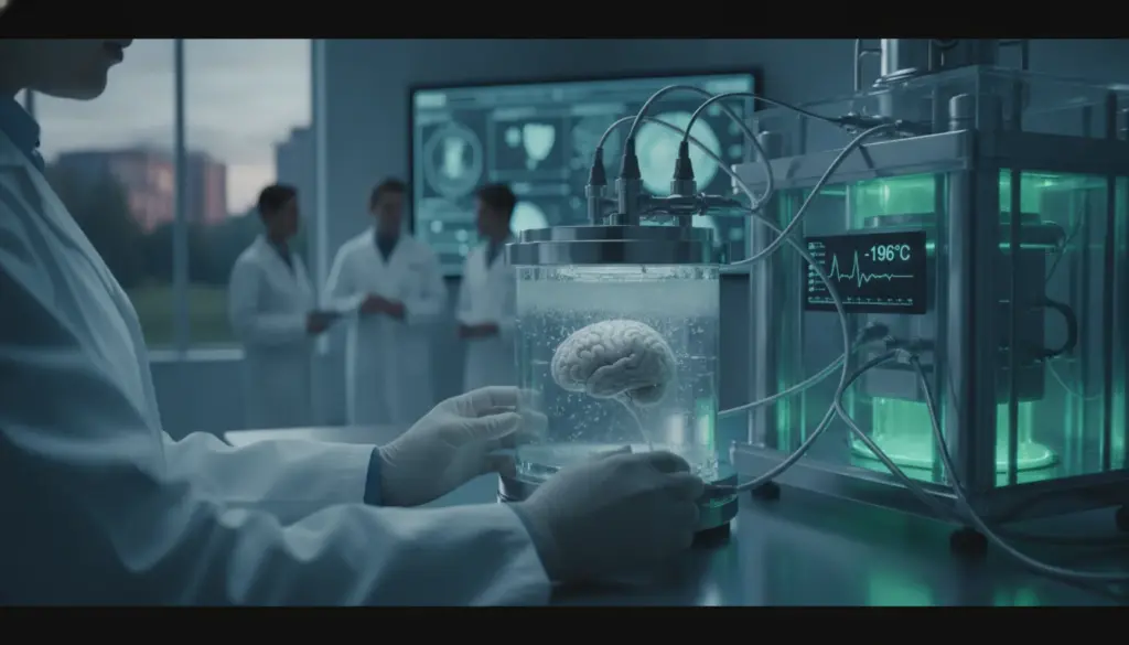
This is exactly where the magic of vitrification comes into play. Instead of allowing crystallization, scientists infuse tissues with specialized cryoprotectants—essentially acting as a biological antifreeze. The biological material is then cooled so rapidly that water molecules simply lack the time to form sharp crystalline structures. Consequently, the tissue solidifies into a smooth, glass-like state, which keeps the intricate cellular architecture completely intact.
From an expert perspective, the most significant hurdle isn’t the plunging temperature itself, but rather the toxic nature of chemical preservatives. The key to this specific trial’s success relied on a unique V3 compound, meticulously designed to minimize chemical degradation while offering robust protection against freezing damage.
Awakening Dormant Neural Pathways
Following the thawing phase, the biological material underwent intense scrutiny. Under an electron microscope, the revived neurons appeared virtually indistinguishable from fresh, unfrozen specimens. They retained their delicate, branching dendrites as well as their mitochondria, the crucial energy-producing centers of the cell. Furthermore, the overall tissue metabolism successfully stabilized, albeit operating at a slightly reduced pace.
The ultimate benchmark, however, was electrical communication. Could these thawed cells still talk to one another? The data revealed a resounding yes. Observers documented long-term synaptic potentiation (LTP), the fundamental mechanism driving how living creatures learn and store memories. This confirms that the organic framework not only endured the deep freeze but held onto its essential data-processing capabilities.
How Different Cells React to Extreme Cold
It is fascinating to note that not all microscopic structures inside the mind handle extreme cold identically. Pyramidal cells located in the CA1 sector required a much stronger electrical nudge to fire up again. In stark contrast, granule cells within the dentate gyrus demonstrated incredible durability, bouncing back to optimal performance almost instantly. Such striking variations highlight the immense complexity of attempting to cryopreserve a diverse, multi-layered organ.
Scaling Up to Complete Organs
Scaling up their ambitions, the team attempted an even bolder experiment: vitrifying an entire brain while it remained inside the skull. They utilized the native vascular network, flushing protective solutions directly through the aorta. Initial attempts hit immediate roadblocks, as the blood-brain barrier and specialized star-shaped astrocytes blocked the fluid, causing severe cellular dehydration.
The scientists overcame this by developing an innovative “alternating equilibrium” technique. This precise protocol allowed them to successfully maintain 70 to 80 percent of the organ’s natural mass alongside its normal shape. While only about a third of these whole-organ trials achieved flawless results, those victorious attempts showcased completely normal neuronal firing upon warming. This represents a monumental leap toward a future where we might see the reliable, long-term storage of life-saving transplant organs.
Current Limitations and Future Realities
Despite the palpable excitement, maintaining a grounded perspective is absolutely necessary. The researchers emphasize that their current methodology is primarily effective on relatively small biological samples. Applying this to a much larger human brain would require highly advanced volumetric heating technologies—such as targeted radio waves—to ensure the warming process is instantaneous and perfectly uniform across deep tissues.
Additionally, the specific genetic impacts of enduring such profound environmental stress remain largely unexplored. Therefore, while these outcomes are incredibly promising, they do not immediately validate human cryonics or the science-fiction dreams of freezing bodies for the distant future. Ultimately, this research validates a profound biological principle: cognitive activity is deeply rooted in physical architecture. If you can perfectly shield the structure, the function will inevitably follow.